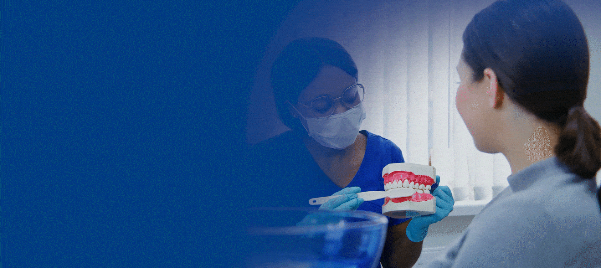
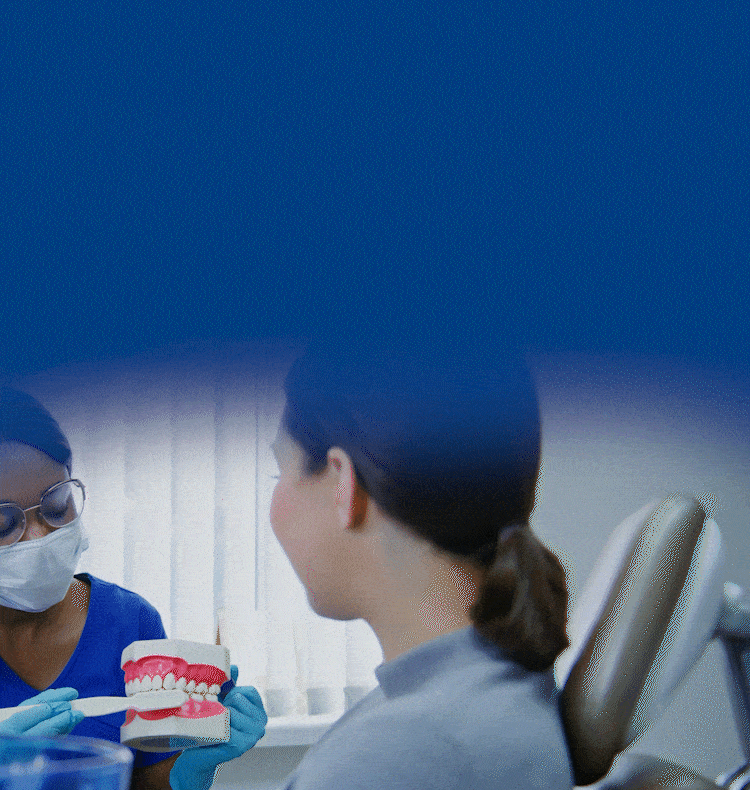
Patients who have positive responses to a brief screening questionnaire will potentially benefit from a clinical examination. A brief clinical examination,8 with the aid of an assistant, should only take a few minutes to complete. The examination and specifications below have been adapted from reliability and validity studies of TMD examinations and diagnostic criteria7 and provide the key information to identify the patient’s clinical diagnoses. This examination collects information about the patient’s pain history, opening and excursive ranges of motion, joint sounds and pain on palpation of the masticatory muscles and TMJs.
Brief Specifications for Conducting the Clinical Examination
The TMD evaluation is primarily based upon history and clinical evaluation, TMJ imaging is only performed if there is a reasonable expectation that the obtained additional information will influence the patient’s treatment.3 In some cases definitive imaging is needed. To determine the actual position of the articular disc, an MRI provides the best diagnostic information. The status of osseous structures such as the condyle, are best viewed with Cone Beam Computed Tomography (CBCT) imaging. A review and report by a radiologist, such as an oral and maxillofacial radiologist, is recommended.
There are other potential assessments that can be used, however, the choice of adjunctive diagnostic procedures should be based upon published, peer-reviewed data showing diagnostic efficacy and safety.9 Additionally, they should only be performed if there is a reasonable expectation that the obtained additional information will influence the patient’s treatment.
Occasionally, but rarely, aggressive local pathology can present with signs and symptoms similar to TMD and all patients who are being evaluated for TMD should receive a careful and comprehensive head and neck assessment.