

The Intraoral and Extraoral Exam
Course Number: 337
Course Contents
Soft Palate and Uvula
This area is examined using direct vision and is normally not palpated unless necessary. If palpation is necessary a topical anesthetic should be used and the tissues should be palpated from the mid line out towards the lateral surfaces. Normally, this area is slightly less vascular than the oropharynx and is usually reddish pink in color (Figure 25e). Observe the area as the patient says “ah.” The tissue should appear loose, mobile and symmetrical during function. The tissue will have a homogenous, spongy consistency on palpation. Atypical observations include yellowish coloring due to increased adipose tissue (especially in older patients), excessively long or short uvulas and uvulas that appear slightly asymmetrical at rest. Occasionally one will discover a bifid (cleft) uvula. Pathologic findings include:
Lesions of any kind
Loss of function or lack of symmetry during function, both of which may indicate compromised swallowing capability and a higher risk of aspiration of food and oral fluids into the lungs
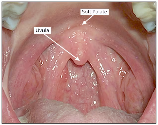
Figure 25e. Soft Palate and Uvula.