

Lasers in Dentistry: Minimally Invasive Instruments for the Modern Practice
Course Number: 394
Course Contents
Flapless Crown Lengthening
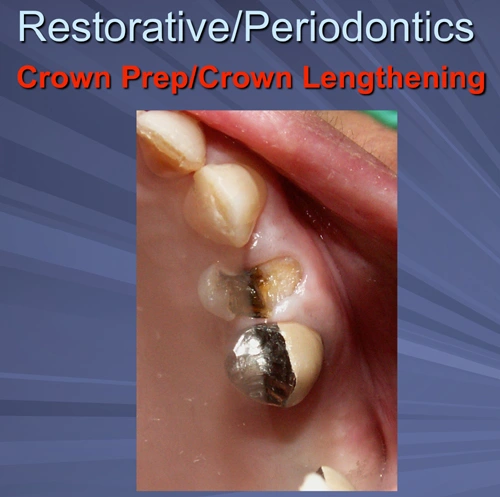
Figure 27. Crown Prep/Crown Lengthening.
Fractured buccal cusp on 70-year-old female requires osseous crown lengthening due to biologic width impingement on mesial and buccal.
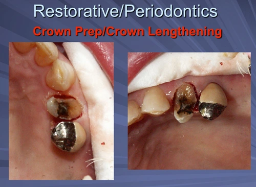
Figure 28. Crown Prep/Crown Lengthening.
The hard tissue crown lengthening can be done with a flapless approach if two millimeters or less of bone needs to be removed. Enough bone has been removed with the Er:YAG laser to assure adequate distance from the final crown margin and osseous crest for biological width requirements.
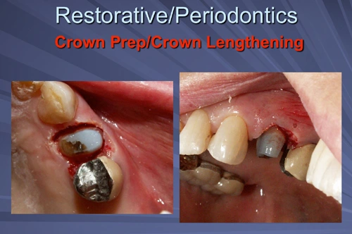
Figure 29. Crown Prep/Crown Lengthening.
The tooth is built up with a high contrast composite material and prepared for the crown on the same appointment as the crown lengthening.
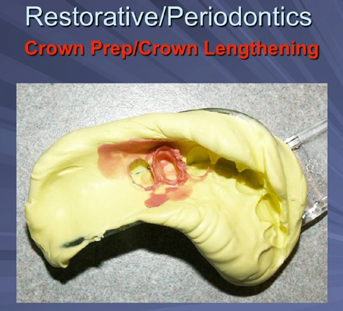
Figure 30. Crown Prep/Crown Lengthening.
A final impression is taken that day as well.
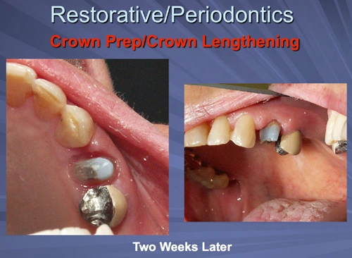
Figure 31. Crown Prep/Crown Lengthening - Two Weeks Later.
The sulcular and attached gingiva are healing well at the crown delivery appointment two weeks after osseous crown lengthening. The patient reported no significant pain post-operatively.
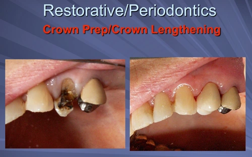
Figure 32. Crown Prep/Crown Lengthening.
The crown is delivered two weeks after the osseous crown lengthening.