

Anomalies of Tooth Structure
Course Number: 651
Course Contents
Dens Evaginatus and Dens Invaginatus
Dens evaginatus is a cusp-like projection of enamel on the tooth crown while dens invaginatus is an inversion or enfolding of enamel into the crown, sometimes extending beyond the CEJ or into the root.7,9 Dens evaginatus is usually found in the central groove or on the lingual ridge of the buccal cusp of a molar or premolar tooth.7,9 Most often, the mandibular premolar teeth are involved bilaterally.7,9 This extra cusp or tubercle is composed of enamel and dentin, and in many instances pulp tissue as well. This particular anomaly occurs in less than 5% of the population, most commonly in Native American, Asian, and aboriginal racial groups.7,9 Exposure and necrosis of the pulp can result from cuspal wear or fracture.9 As discussed, dens evaginatus is an external outcropping of tooth structure in contrast to dens invaginatus, an internal involution of tooth structure.7,9
Dens invaginatus varies in the degree of tooth structure enfolding. Three types are recognized per Oehlers Classification, the most widely used nomenclature (Table 4).10,11 These coronal invaginations usually involve the permanent maxillary lateral incisors but other permanent teeth can be affected including central incisors, premolar, canine, and molar teeth with the maxillary arch more frequently involved than the mandibular arch.7,9,10 The least severe form, dens invaginatus, typically involves the cingulum on the lingual surface of the crown of permanent maxillary lateral incisors and may appear as a small pit (Figure 32).7,9 A more severe coronal type of invagination, dens in dente or tooth within a tooth, arises from the incisal edge of the involved tooth (Figure 33).7,9 The permanent maxillary lateral incisor teeth are most commonly affected with a tendency for bilateral occurrence.9 The most severe form, dilated odontome, involves the tooth root with a doughnut-shaped invaginated defect lined with cementum.7,9 Radiographically, the affected tooth is significantly malformed displaying a radiopaque round or oval periphery with a radiolucent center as seen in Figure 34.7,9,12 This anomaly is rare and appears to be independent of the traditional classification system outlined in Table 4.12 The clinical relevance of these defects is the possibility of pulpal involvement and necrosis.7,9,10
| Type | Description |
|---|---|
| Type I | Defect is enamel-lined and confined to the tooth crown |
| Type II | Defect is enamel-lined and extends into the pulp chamber but remains within the root canal |
| Type III | Defect extends into the tooth root and perforates the root laterally or through the apical foramen (See subtypes A and B) |
| Type IIIA | Perforates the root laterally with PDLS communication; ususally without pulp involvement |
| Type IIIB | Communicates with the PDLS at the apical foramen; ususally without pulp involvement |
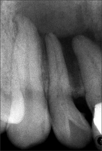
Figure 32.
Periapical radiograph of tooth #7 with dens invaginatus Type I.
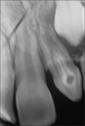
Figure 33.
Periapical radiograph of tooth #10 with dens invaginatus Type II.
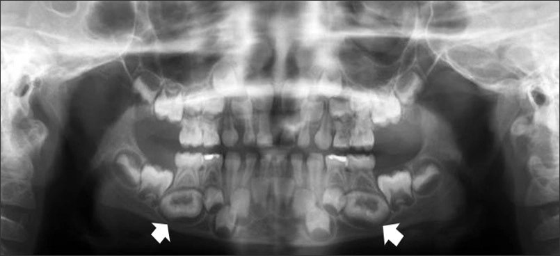
Figure 34.
Panoramic radiograph with developing dilated odontomes of mandibular premolar teeth #20 & #29.