

A Guide to Clinical Differential Diagnosis of Oral Mucosal Lesions
Course Number: 110
Course Contents
Benign Mesenchymal Tumors of Oral Mucosa
For purposes of this discussion, mesenchymal tumors are composed of fibrous connective tissue, smooth muscle, skeletal muscle, blood and lymphatic vessels, adipose tissue, and peripheral nerve tissue. Unless otherwise noted in the following descriptions, benign mesenchymal tumors clinically present as well-circumscribed, persistent, slowly growing, non-tender, soft tissue enlargements.
Irritation fibroma, epulis fissuratum, and peripheral ossifying fibroma represent an overgrowth of fibrous connective tissue.
Irritation fibroma* is a common reactive soft tissue enlargement due to chronic irritation or trauma. It most commonly presents as an exophytic, dome-shaped enlargement which may be firm or compressible to palpation. The mucosa overlying the lesion may be normal or ulcerated due to trauma. It is most common on the buccal and labial mucosa. Treatment for irritation fibroma is excisional biopsy and microscopic diagnosis. Recurrence is uncommon.
Irritation fibroma

Irritation fibroma
Epulis fissuratum (inflammatory fibrous hyperplasia)* represents hyperplasia of dense connective tissue due to chronic irritation from the flange of a denture. It appears as an enlargement in the vestibule. Often a fissure will be present in the lesion, corresponding to the location of the denture flange. Treatment consists of surgical excision of the mass and microscopic diagnosis, and usually remaking or relining the denture.
Epulis fissuratum

Epulis fissuratum
Peripheral ossifying fibroma (peripheral fibroma)*, is a reactive soft tissue enlargement arising from cells of the periodontal ligament. It is always located on the gingiva or attached alveolar mucosa, often ulcerated, and may be red or have a normal mucosal color. It is most common in adolescents through young adults. An interesting feature microscopically is that peripheral ossifying fibroma frequently forms a mineralized product within a cellular fibrous stroma. Treatment is excisional biopsy and microscopic diagnosis. The lesion should be removed down to periosteum. It has a good prognosis although recurrence rates up to 16% have been reported. Treatment for recurrent lesions is re-excision.
Peripheral ossifying fibroma

Peripheral ossifying fibroma
Schwannoma* is a benign neoplasm of Schwann cells. It is firm, encapsulated, and often freely moveable. Treatment is excisional biopsy and microscopic diagnosis. Recurrence is uncommon.
Schwannoma

Schwannoma
Neurofibroma is also a benign neoplasm of Schwann cells. Neurofibroma most commonly occurs as a solitary lesion, but multiple neurofibromas are a characteristic feature of neurofibromatosis. Solitary neurofibroma is fixed to surrounding structures and may be firm or compressible upon palpation. Treatment consists of excisional biopsy and microscopic diagnosis. Recurrence is not expected.
Traumatic (or amputation) neuroma* represents a reactive proliferation of nerve bundles following severing of a nerve. It arises most commonly in locations containing relatively large peripheral nerves, such as the mental foramen, tongue and lower lip. Neuroma is often, but not always, painful to palpation. Multiple neuromas unassociated with trauma are part of multiple endocrine neoplasia type 2B syndrome. Treatment consists of excisional biopsy and microscopic diagnosis. Lesions usually do not recur.
Granular cell tumor* is a benign neoplasm previously called granular cell myoblastoma. The tumor cells are of Schwann cell origin. The lesion is fixed to surrounding structures. The most common location is the dorsum of the tongue. Microscopically, the lesion often appears infiltrative, however, conservative excision and microscopic diagnosis is usually curative.
Granular cell tumor

Granular cell tumor
Rhabdomyoma of the oral mucosa is a rare benign neoplasm of skeletal muscle origin. It is located only where skeletal muscle is found. The most common location is the tongue. It is fixed to surrounding structures. Treatment is excisional biopsy and microscopic diagnosis.
Congenital epulis* is a benign soft tissue enlargement that occurs on the attached alveolar mucosa of infants. Almost 90% of these lesions occur in females. Treatment is surgical excision and microscopic diagnosis. Prognosis is excellent.
Congenital epulis

Congenital epulis
The following benign mesenchymal tumors have clinical features of vascular lesions: peripheral giant cell granuloma, pyogenic granuloma, hemangioma, leiomyoma, and sometimes peripheral ossifying fibroma as discussed above. A vascular soft tissue enlargement is red, blue, or purple and blanches upon pressure.
Peripheral giant cell granuloma* is a reactive soft tissue enlargement that occurs only on gingiva or attached alveolar mucosa. Treatment is excisional biopsy and microscopic diagnosis. The microscopic features consist of giant cells that are identical to those of central giant cell granuloma.
Peripheral giant cell granuloma

Peripheral giant cell granuloma
Pyogenic granuloma* is a soft tissue enlargement that develops in reaction to minor injury or irritation. It can be found on any oral mucosal surface at any age, but is most common on the gingiva in children and pregnant females. Pyogenic granuloma is compressible, can be lobulated and is often pedunculated. Ulceration is frequently present. The initial growth rate is quite rapid. In lesions of longer duration collagen replaces much of the vascularity and the lesion begins to resemble an irritation fibroma. Treatment is excisional biopsy and microscopic diagnosis. Recurrence is not unusual, and recurrent lesions should be re-excised. For pregnant patients excision can be deferred until after the pregnancy is completed.
Pyogenic granuloma

Pyogenic granuloma
Hemangioma* is a proliferation of blood vessels which usually is noted at birth or early childhood. It may be well circumscribed or diffuse. The arteriovenous malformation is a different lesion. It represents a direct communication between an artery and a vein, and it will demonstrate a thrill and bruit. A hemangioma requires no treatment unless it is a functional or cosmetic problem. Many hemangiomas will regress spontaneously during childhood. Incision of an arteriovenous malformation may lead to fatal hemorrhage.
Hemangioma

Hemangioma
Leiomyoma is a benign neoplasm of smooth muscle. In the oral cavity it arises from smooth muscle in the wall of blood vessels. It is firm and sometimes has a vascular appearance. Treatment is excisional biopsy and microscopic diagnosis. It does not tend to recur.
Lymphangioma is a developmental overgrowth of lymphatic vessels and not a true neoplasm. In almost all cases lymphangioma is present at birth or appears during the first 2 years of life. The most common locations are the neck and the tongue. Tongue lesions can cause macroglossia, leading to problems with eating and speaking. Tongue lesions are usually compressible and fixed to surrounding structures. The mucosa overlying the lesion often has multiple nodules resembling small vesicles. Lymphangiomas are unlikely to undergo spontaneous regression. Surgical removal of the lesion is difficult if the lesion is poorly circumscribed. The prognosis for most patients is good, but occasionally lymphangioma can cause airway obstruction and be life-threatening.
To view the Decision Tree for Oral Mucosal Lesions, click on one of the options shown.
 View Interactive
 View as PDF
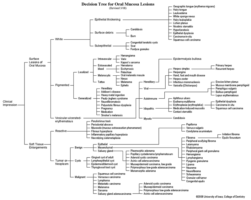 View as GIF
To view the Decision Tree for Oral Mucosal Lesions, click on one of the options shown.