

A Guide to Clinical Differential Diagnosis of Oral Mucosal Lesions
Course Number: 110
Course Contents
Localized Pigmented Surface Lesions of Oral Mucosa
Localized pigmented surface lesions are divided into 4 categories based on their cause and clinical features.
Intravascular Blood Lesions appear red, blue or purple due to an increased amount of blood within blood vessels as a result of increased number or size of blood vessels. Firm palpation of the lesions causes them to blanch because the blood is displaced.
Hemangioma* is a proliferation of blood vessels which usually is noted at birth or early childhood. It may be well circumscribed or diffuse. The arteriovenous malformation is a different lesion. It represents a direct communication between an artery and a vein, and it will demonstrate a thrill and bruit. A hemangioma requires no treatment unless it is a functional or cosmetic problem. Many hemangiomas will regress spontaneously during childhood. Incision of an arteriovenous malformation may lead to fatal hemorrhage.
Hemangioma

Hemangioma
A varix* is a dilated vein or venule. It presents as a relatively small, localized, elevated, blue or purple lesion. It is compressible and blanches upon pressure unless a thrombus has formed within it. A thrombosed varix is firm and does not blanch. Varices are most common on the ventral surface of the tongue, floor of the mouth, lips, and buccal mucosa. Varices increase in number with age and may also be the result of trauma. Once a varix has been diagnosed it needs no further treatment. A thrombosed varix often cannot be clinically distinguished from a nevus, and biopsy and microscopic examination are necessary to establish a definitive diagnosis.
Varix

Varix
Kaposi sarcoma* is a malignant vascular neoplasm most commonly seen in patients with HIV infection, organ transplants or other causes of immune suppression. It appears as a flat or slightly elevated, blue to purple plaque on skin and oral mucosa. The lesion may develop into a compressible soft tissue enlargement that sometimes blanches on pressure. Oral lesions occur most commonly on the hard palate and gingiva. Treatment for oral Kaposi sarcoma includes systemic or intralesional chemotherapy and surgical excision. Prognosis depends on the systemic health status of the patient.
Kaposi sarcoma

Kaposi sarcoma
To view the Decision Tree for Oral Mucosal Lesions, click on one of the options shown.
 View Interactive
 View as PDF
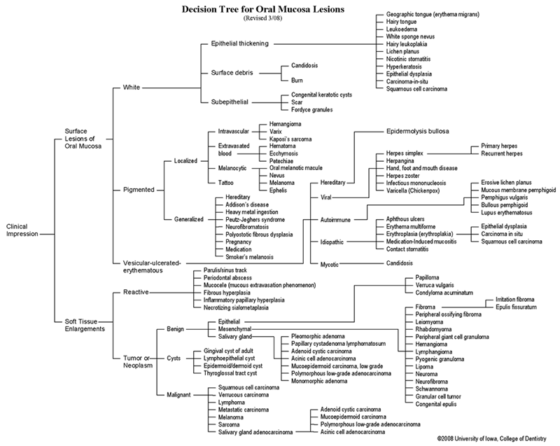 View as GIF
To view the Decision Tree for Oral Mucosal Lesions, click on one of the options shown.