

A Guide to Clinical Differential Diagnosis of Oral Mucosal Lesions
Course Number: 110
Course Contents
Tattoo
Tattoo* is a localized pigmented area caused by implantation of foreign material into skin or oral mucosa. Oral tattoos are usually caused by amalgam particles or graphite in lead pencils. A tattoo is localized, dark gray to black, non-tender, and either macular or slightly thickened. A tattoo sometimes increases in size due to ingestion of the foreign material by phagocytes and then migration of these cells. Some tattoos can be visualized on a radiograph, but absence of radiographic evidence of amalgam particles does not exclude the diagnosis of tattoo. Obviously, some tattoos are intentional artistic endeavors and do not cause a diagnostic challenge.
The typical small, localized, non-thickened tattoo does not require treatment, once a diagnosis is made. A tattoo that is thickened and does not have amalgam particles evident on a radiograph should be biopsied so that nevus and melanoma can be excluded.
Tattoo

Tattoo
To view the Decision Tree for Oral Mucosal Lesions, click on one of the options shown.
 View Interactive
 View as PDF
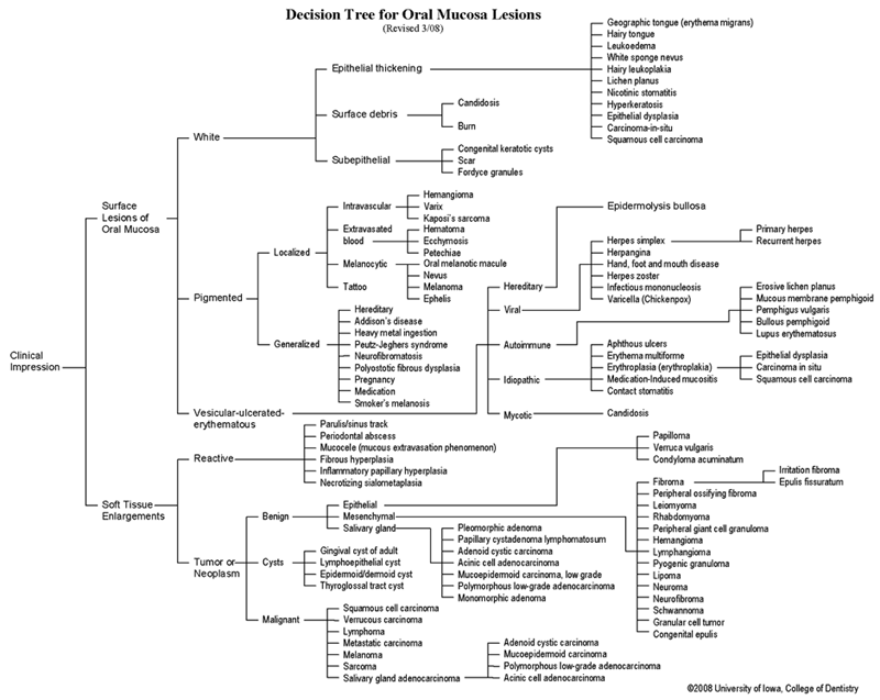 View as GIF
To view the Decision Tree for Oral Mucosal Lesions, click on one of the options shown.