

A Guide to Clinical Differential Diagnosis of Oral Mucosal Lesions
Course Number: 110
Course Contents
White Surface Lesions
Erythema migrans (geographic tongue, benign migratory glossitis) is a common, harmless lesion that can typically be diagnosed by its clinical features. It presents as multiple red patches surrounded by a thickened, irregular, white border. A lesion will resolve in one area and appear in other areas (migrate). This condition is usually not painful and requires no treatment. If the patient complains of pain or burning with the lesions, a diagnosis of concomitant candidosis or burning mouth syndrome should be considered. Rarely, lesions of erythema migrans can be found on oral mucosal surfaces other than the tongue.
Erythema migrans

Erythema migrans
Nicotine stomatitis* is an epithelial thickening lesion of the hard palate caused by heat from smoking a pipe, cigar, or occasionally cigarettes. The lesion is white, rough, asymptomatic, and leathery appearing and contains numerous red dots or macules. The red macules represent inflamed salivary gland duct orifices. Nicotine stomatitis is not considered a premalignant lesion and does not need to be biopsied. However, the patient should be encouraged to stop smoking, and the oral mucosa should be evaluated periodically. The prognosis for nicotine stomatitis is good, but the patient is at increased risk to develop cancer in other locations in the upper aerodigestive tract.
Nicotine stomatitis

Nicotine stomatitis
White sponge nevus* is a genetic disorder, usually congenital or developing in childhood. The oral mucosa is diffusely white, rough, thickened and folded. The most common location is the buccal mucosa bilaterally, but other oral mucosal areas may be involved. Nasal, pharyngeal, and anogenital mucosa may be affected. The condition is not painful. Other family members often have the condition. The clinical features and history are diagnostic. This condition is benign and requires no treatment. The prognosis is excellent.
White sponge nevus

White sponge nevus
Leukoedema is a generalized white change of oral mucosa which is probably a variation of normal rather than a disease. The cause is unknown. It occurs much more commonly in black people than white people. Leukoedema is diffuse and symmetrically distributed on the buccal mucosa and may extend onto the labial mucosa. The appearance is gray-white, opaque, or milky. It can be smooth to palpation or wrinkled, and it does not rub off. A characteristic clinical feature is that the white appearance decreases when the buccal mucosa is stretched. Leukoedema is asymptomatic, and the patient is unaware of its presence. Leukoedema is diagnosed clinically, and a biopsy is not required. No treatment is necessary. It is a benign lesion and is not premalignant.
Lichen planus* is a chronic inflammatory disease involving skin and oral mucosa. It represents an immune abnormality involving T lymphocytes sensitized to antigens in the overlying stratified squamous epithelium. Often it is associated with medications the patient is taking, and it is then called a lichenoid mucositis secondary to medications. Classic lichen planus and drug-related lichenoid mucositis appear identical clinically and microscopically.
Lichen planus

Lichen planus
Skin lesions of lichen planus consist of pruritic (itching), erythematous to light purple patches, sometimes with an overlying network pattern of white lines or striations. Oral lesions most commonly appear as white epithelial thickening arranged in a network pattern (Wickham striae) with erythema of the surrounding mucosa. White patches, erythematous erosions, and ulcers may also occur. The white lesions are not painful, but the erosions and ulcers are usually painful. Lichen planus almost always has multiple lesions bilaterally, with the buccal mucosa commonly involved. Oral lesions may occur with or without skin lesions.

Skin lesions of lichen planus
Asymptomatic lesions require no treatment other than inspection during annual dental visits. Topical and/or systemic corticosteroids will almost always control, but not cure, painful erosions and ulcers of lichen planus. If suspected lichen planus is refractory to traditional treatment, an incisional biopsy may be required for definitive diagnosis.
The term leukoplakia refers to a clinically white mucosal thickening lesion that cannot be further defined. Most “leukoplakia” will be shown microscopically to be hyperkeratosis, with or without epithelial dysplasia, carcinoma in situ, or superficially invasive squamous cell carcinoma. Leukoplakia is a clinical description—not a diagnosis, and the term will not be used further in this discussion.
Hyperkeratosis (focal keratosis)* is a microscopic term meaning increased thickness of the keratin layer of stratified squamous epithelium with no microscopic evidence of atypical epithelial cells. Clinically, hyperkeratotic lesions appear as white, rough, non-painful patches that do not rub off. They are often secondary to chronic irritation, such as biting or tobacco use.
Hyperkeratotic lesions on oral mucosal surfaces that are normally keratinized, such as dorsum of the tongue, hard palate, and attached gingiva, sometimes represent a physiologic response (callus) to chronic irritation. These lesions will usually resolve if the irritant is removed. Hyperkeratotic lesions on surfaces that are normally nonkeratinized are potentially more serious and should be biopsied if they do not resolve if irritants are removed. Remember, however, that dysplasia, carcinoma in situ, and squamous cell carcinoma can occur on any oral mucosal surface.
Hyperkeratosis

Hyperkeratosis
Epithelial dysplasia is atypical or abnormal growth of the stratified squamous epithelium lining a mucosal surface. It is a diagnosis that must be made microscopically. These lesions appear clinically as white, rough, non-painful areas, or non-painful red patches (“erythroplakia” or “erythroplasia”), or patches that demonstrate both red and white areas. Because these lesions are asymptomatic, the patient is usually not aware of them. Some lesions diagnosed as epithelial dysplasia will progress to squamous cell carcinoma, while others will resolve. Since it is impossible to determine by microscopic examination which lesions will progress or resolve, treatment is complete surgical excision, if possible, and follow-up.
Carcinoma in situ* is cancer of the oral epithelium which is confined to the epithelial layer. It presents most commonly as a persistent red plaque (erythroplakia) or a mixed white and red plaque. It may also appear as a white plaque. Complete removal is the treatment. When completely removed, the prognosis is excellent, although the patient is at increased risk of developing new lesions at other locations on the oral mucosa.
Carcinoma in situ

Carcinoma in situ
Squamous cell carcinoma* is the most common malignant neoplasm of the oral cavity. Tobacco and alcohol are the most common risk factors, but squamous cell carcinoma can occur in patients with no known risk factors. Squamous cell carcinoma can occur anywhere on the oral mucosa, but is most common on the ventral and lateral surfaces of the tongue, floor of the mouth, soft palate, tonsillar pillar area, and retromolar trigone areas.
Superficially invasive, or early, squamous cell carcinoma lesions appear as surface lesions rather than soft tissue enlargements. They are almost invariably non-painful, and thus patients do not know they have a lesion. Early lesions may be white rough epithelial thickening lesions, red persistent non-painful lesions, or a combination of the two.
It is important to recognize squamous cell carcinoma in its early stages when cure is possible without disfiguring surgery. The main treatment for oral squamous cell carcinoma is complete surgical excision. Lymph node dissection is performed when lymph nodes are involved. Radiation therapy is often used as an adjunct to surgery. Chemotherapy is reserved for palliative therapy.


Squamous cell carcinoma
To view the Decision Tree for Oral Mucosal Lesions, click on one of the options shown.
 View Interactive
 View as PDF
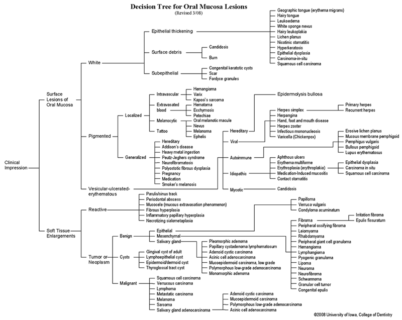 View as GIF
To view the Decision Tree for Oral Mucosal Lesions, click on one of the options shown.