

A Guide to Clinical Differential Diagnosis of Oral Mucosal Lesions
Course Number: 110
Course Contents
Malignant Neoplasms of Oral Mucosa
A challenge in differential diagnosis of soft tissue enlargements is the distinction between malignant and reactive lesions. Both can be rapidly growing and painful. The key distinction is that malignant neoplasms are persistent and progressive, while reactive lesions fluctuate in size or eventually regress. Reactive lesions may be associated with soft, tender lymph nodes, while lymph nodes involved with metastatic malignant neoplasms are firm and non-tender.
Squamous cell carcinoma* is the most common malignant neoplasm of the oral cavity. Tobacco and alcohol use have been identified as risk factors, but squamous cell carcinoma can occur in patients with no known risk factors. This tumor can occur anywhere on the oral mucosa, but it is most common on the ventral and lateral surfaces of the tongue, floor of the mouth, soft palate, tonsillar pillar area, and retromolar trigone. Advanced squamous cell carcinoma presents as an indurated (hard) tumor mass fixed to surrounding structures. It is often ulcerated and may be painful. It may be associated with cervical lymphadenopathy representing metastatic lesions. Early squamous cell carcinoma and its precursor lesions are almost invariably asymptomatic, and thus patients do not know they have a lesion. Early lesions may be white rough epithelial thickening lesions (leukoplakia), red persistent non-painful lesions (erythroplakia) or a combination of the two. It is important to discover squamous cell carcinoma in its early stages when cure is possible without disfiguring surgery. The main treatment is complete surgical excision. Lymph node dissection is performed when lymphadenopathy is evident. Radiation therapy is often used as an adjunct to surgery. Chemotherapy is reserved for palliative therapy. Verrucous carcinoma is a slowly-growing, low-grade variation of squamous cell carcinoma. The lesion has a rough warty surface and is usually asymptomatic. Verrucous carcinoma can invade underlying tissue but almost never metastasizes. It has a good prognosis compared to typical oral squamous cell carcinoma.
Salivary gland adenocarcinoma includes polymorphous low-grade adenocarcinoma, adenoid cystic carcinoma, acinic cell adenocarcinoma, mucoepidermoid carcinoma, carcinoma arising in pleomorphic adenoma, and a number of other lesions. These lesions may grow rapidly or slowly and present with pain and paresthesia or be asymptomatic. They all demonstrate infiltrative growth. Treatment is generally complete surgical excision. Prognosis depends upon the stage or extent of the tumor and its microscopic features.
Salivary gland adenocarcinoma

Salivary gland adenocarcinoma
Lymphomas* are a diverse group of malignant neoplasms of lymphocytes and their precursors. They form solid tumor masses and usually arise within lymphoid tissue. Lymphomas are subdivided into Hodgkin disease and non-Hodgkin lymphomas. The most common presentation of Hodgkin's disease in the head and neck area is persistent, progressive enlargement of cervical and supraclavicular lymph nodes. Hodgkin disease only rarely has intraoral lesions. Non-Hodgkin lymphomas include numerous different lesions that may arise in lymph nodes or in extranodal sites. Lesions arising in lymph nodes are non-tender, slowly enlarging masses that eventually become multiple fixed enlargements. Extranodal lymphoma in the oral cavity may be the first manifestation of lymphoma or may be part of a disseminated process. Extranodal oral lymphoma of soft tissue is typically a non-tender, poorly circumscribed, compressible, soft tissue enlargement, sometimes with erythema and ulceration of the overlying mucosa. The most common sites are Waldeyer ring, posterior hard palate, buccal mucosa, or gingiva. Lesions may also arise within the jaws. Jaw lesions have clinical features similar to other malignancies of bone. Malaise, fever, and weight loss may accompany both Hodgkin disease and non-Hodgkin lymphoma. The management of lymphoma involves biopsy of the lesion to obtain a definitive diagnosis. This is followed by staging to determine the extent of the disease. Chemotherapy and/or radiation therapy are used for treatment. The prognosis is extremely variable.
Lymphomas

Lymphomas
Carcinomas metastatic to oral soft tissue: Metastatic neoplasms to the oral cavity make up only 1% of all oral cancers, and these tumors are found much more frequently in the bone of the jaws than in the oral soft tissues. The vast majority of tumors that metastasize to the oral cavity are adenocarcinomas. The most common primary locations of these tumors include breast, lung, kidney, gastrointestinal tract (stomach and colon), thyroid and prostate.
The most common oral mucosal locations for metastatic carcinoma are the gingiva and tongue. Early lesions are dome-shaped nodules with a smooth, normal-appearing surface. These lesions may appear benign clinically. Later, the surface may become ulcerated and necrotic, and the lesion may bleed easily. These lesions appear malignant clinically.
Sarcomas are relatively rare malignant neoplasms of non-epithelial tissue. Sarcomas may arise in soft tissue or bone. Examples include fibrosarcoma, rhabdomyosarcoma (skeletal muscle origin), and leiomyosarcoma (smooth muscle origin). Sarcomas generally are rapidly growing, poorly circumscribed, infiltrative, and cause ulceration of the overlying tissue. Treatment is usually surgical removal combined with chemotherapy and/or radiation therapy. The prognosis depends upon the stage of the disease and microscopic features.
Melanomas are relatively rare in the oral cavity. They are discussed in the section on localized pigmented surface lesions.
To view the Decision Tree for Oral Mucosal Lesions, click on one of the options shown.
 View Interactive
 View as PDF
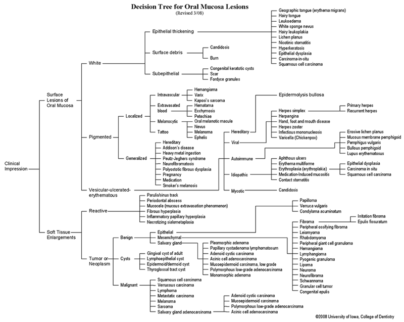 View as GIF
To view the Decision Tree for Oral Mucosal Lesions, click on one of the options shown.