

A Guide to Clinical Differential Diagnosis of Oral Mucosal Lesions
Course Number: 110
Course Contents
How to Use the Decision Tree
To use the decision tree, the clinician begins at the left side of the tree, makes the first decision, and proceeds to the right. The names of individual lesions are listed on the far right of the tree. Any lesion or group of lesions that cannot be excluded becomes part of the clinical differential diagnosis.
The first decision to make when using the decision tree is whether the lesion is a surface lesion or soft tissue enlargement.
Surface lesions consist of lesions that involve the epithelium and superficial connective tissue of mucosa and skin. They do not exceed 2-3 mm in thickness. Surface lesions are divided into three categories based on their clinical appearance: white, pigmented, and vesicular-ulcerated-erythematous. Each of these categories is further subclassified as shown in Tables 1-3.
Table 1. White Surface Lesions of Oral Mucosa.
| EPITHELIAL THICKENING WHITE LESIONS Asymptomatic; rough to palpation; fixed to the surface (won’t rub off) | |
|---|---|
| Lichen planus | Multiple areas of mucosa involved; bilateral distribution; white plaques arranged in striated pattern associated with erythema; ulcers may be present; skin lesions may be present. |
| Nicotine (nicotinic) stomatitis | Hard palate; mainly in pipe or cigar smokers. |
| Hairy tongue | Dorsum of tongue. |
| Hairy leukoplakia | Lateral surface of tongue; patient is immunocompromised, e.g. AIDS. |
| White sponge nevus | Multiple lesions affecting broad areas of mucosa; familial history; present from early age; genital & rectal mucosa may be affected. |
| Leukoedema | Bilateral on buccal mucosa. Disappears when tissue is stretched. |
| Erythema migrans (geographic tongue, benign migratory glossitis) | Multiple red patches with irregular yellow-white border; dorsal lateral tongue; lesions migrate; usually asymptomatic. |
| Hyperkeratosis | May resolve spontaneously. |
| Epithelial dysplasia Carcinoma-in-situ Superficially invasive squamous cell carcinoma | Persistent; usually asymptomatic; more common as red lesion or mixed red and white lesion. |
| SURFACE DEBRIS WHITE LESIONS Pain or burning; rubs off; submucosal erythema | |
| Candidosis | History of antibiotic therapy, immunosuppression; xerostomia; nail and/or vaginal lesions may be present |
| Burn (thermal or chemical) | History of burn. |
| Dried, thick saliva | Removed with wet gauze. |
| SUBEPITHELIAL WHITE LESIONS Asymptomatic; smooth to palpation; surface is translucent. | |
| Cysts | Small cysts of oral mucosa can appear white. Examples are congenital keratotic cyst and lymphoepithelial cyst. |
| Fordyce granules (ectopic sebaceous glands) | Yellow, circumscribed, in clusters; most commonly located on buccal mucosa and upper lip. |
| Mucosal scarring | History of injury or surgery; usually poorly defined. |
Table 2. Localized Pigmented Surface Lesions of Oral Mucosa.
| INTRAVASCULAR BLOOD LESIONS Usually blanch on pressure and compressible | |
|---|---|
| Varix | Blue; thickened; sometimes does not blanch due to thrombosis. |
| Hemangioma | Congenital; thickened; red or blue |
| Kaposi sarcoma | Patient is immunocompromised; may be thickened or flat. |
| EXTRAVASCULAR BLOOD LESIONS Do not blanch; present for less than 1 month; may have history of injury or bleeding problem. | |
| Hematoma | Thickened; firm to palpation. |
| Ecchymosis | Not thickened |
| Petechiae | Focal and pinpoint size; red; multiple; not thickened |
| MELANOCYTIC LESIONS Persistent; do not blanch | |
| Ephelis (freckle) | Not thickened; located on sun-exposed surfaces. |
| Oral melanotic macule | Not thickened; located on mucosa not exposed to sun |
| Nevus | Thickened; may be flat early in development |
| Melanoma | Thickened; may be flat early in development |
| TATTOO Do not blanch; may be history of injury; radiopaque object sometimes seen on radiograph; may be thickened or flat. | |
Table 3. Vesicular-Ulcerated-Erythematous Surface Lesions of Oral Mucosa.
| HEREDITARY – EPIDERMOLYSIS BULLOSA Skin lesions are always present; Nikolsky sign often present; mouth opening may be restricted due to scarring. Lesions are congenital or begin at an early age; patient frequently has a familial history. | |
|---|---|
| MYCOTIC – CANDIDOSISv(CANDIDIASIS) Diffuse mucosal erythema; burning or pain may be present; ulcers are rarely present; lymphadenopathy is rare. Patient often has predisposing factors: antibiotics, immunosuppression. | |
| AUTOIMMUNE Slow onset; chronic lesions; exacerbations & partial remissions; lesions do not heal in a predictable period of time; lymphadenopathy is rare. | |
| Mucous membrane pemphigoid (cicatricial pemphigoid; benign mucous membrane pemphigoid) | Erythematous attached gingiva; vesicles sometimes observed; Nikolsky sign may be present; skin vesicles & ulcers may be present. |
| Bullous pemphigoid | Skin vesicles, bullae & ulcers are always present; occasional oral vesicles & ulcers. |
| Pemphigus | Mucosal vesicles & ulcers in any location usually precede skin lesions; Nikolsky sign may be present. |
| Lupus erythematosus | Nonspecific mucositis & ulcers are sometimes present but are associated with skin lesions. Oral lesions: white epithelial striae with submucosal erythema (lichenoid lesions). Multiple organ system disorders: erythematous skin rash, photosensitivity, arthritis, nephritis, neurologic disease; anemia, leukopenia, thrombocytopenia. |
| VIRAL Acute onset; multiple lesions; systemic manifestations (malaise, fever, diarrhea, lymphadenopathy, lymphocytosis) often present; vesicle stage is present in all except mononucleosis. | |
| Herpes simplex virus: | |
| Primary herpes | Vesicles & ulcers may be present anywhere in the oral cavity, pharynx, lips or perioral skin; gingiva is edematous & erythematous; lymphadenopathy is common; malaise, fever & diarrhea in some cases. |
| Recurrent herpes | Occurs on sun-exposed surfaces of lips; intraorally occurs on keratinized mucosa (dorsum of tongue, hard palate, attached gingiva); usually recurs in same location; heals in a predictable period of time for each patient. |
| Varicella (chickenpox) | Crops of pruritic papules, vesicles, ulcers on trunk spreading to arms, legs & face; mild malaise, fever & lymphadenopathy; occasional oral ulcers. |
| Herpes zoster (shingles) | Prodromal pain followed by vesicles & ulcers in the distribution of a sensory nerve; unilateral lesions; postherpetic neuralgia may occur. |
| Herpangina (Coxsackievirus A) | Vesicles & ulcers in posterior oral cavity & pharynx; may have mild systemic manifestations. |
| Hand, foot and mouth disease (Coxsackievirus A) | Vesicles & ulcers of oral & pharyngeal mucosa; vesicles & macules on hands and feet; mild systemic manifestations. |
| Rubeola (measles) | Fever, conjunctivitis, photophobia, cough, nasal discharge; oral vesicles (Koplik spots); erythematous maculopapular skin rash on face spreading to trunk & extremities. |
| Epstein-Barr virus | Infectious mononucleosis Generalized lymphadenopathy; splenomegaly; hepatomegaly; palatal petechiae; erythematous oral & pharyngeal mucosa; occasionally mucosal ulcers; no vesicular stage. |
| IDIOPATHIC Each disease must be considered as a separate entity. | |
| Aphthous ulcers | Abrupt onset of recurrent ulcers on nonkeratinized mucosal surfaces; individual ulcers heal in a predictable period of time which is variable for each patient; may be menstrually related; familial history common; “herpetiform” aphthae refer to multiple crops of small aphthous ulcers; “major” aphthae are deeper, longer lasting and more frequent ulcers which often heal with scarring. |
| Erosive lichen planus | Erythematous mucosal lesions usually with areas of ulceration; often bilateral distribution; white epithelial striae at edge of erythematous areas; atrophy of filiform papillae may be seen; chronic course. |
| Medication-induced mucositis | A variety of drugs cause mucosal lesions that do not appear to be allergic in nature; mucosal lesions consists of ulcers and erosions occurring on both keratinized & nonkeratinized mucosal surfaces. |
| Contact stomatitis | Burning, pain, ulcers, erosions, erythema sometimes covered with shaggy hyperkeratosis. Most commonly secondary to cinnamon flavoring. |
| Erythema multiforme | Sudden onset of diffuse mucosal ulcers involving buccal & labial mucosa; sometimes recurrent with variable periods of remission; skin lesions present “iris” or “target” appearance on palmar & plantar surfaces; lymphadenopathy is rare. |
| Erythroplasia (erythroplakia): epithelial dysplasia, carcinoma in situ, superficially-invasive squamous cell carcinoma | Asymptomatic, persistent, erythematous, velvety, focal to diffuse mucosal areas; more common in heavy consumers of alcohol. |
Soft tissue enlargements are swellings or masses that are divided into two categories: reactive and tumors in Table 4. The term tumor is used in the clinical sense of an enlargement and is not based on microscopic criteria or basic pathologic process. For example, irritation fibroma is classified as a tumor because this lesion is persistent and progressively increases in size, although most people agree that the true pathogenesis is that of a reactive process secondary to chronic irritation.
Table 4. Soft Tissue Enlargements.
| Reactive | Tumors |
|---|---|
| Regress, resolve | Persistent and progressive |
| Often symptomatic | Often asymptomatic |
| Growth rate: hours, days, weeks | Growth rate: weeks, months, years |
| Fluctuate in size | Persistent & progressive |
| Sometimes associated with tender, soft lymph nodes | Lymph nodes not enlarged unless associated with metastatic cancer; then they are firm & non-tender |
| Sometimes associated with systemic manifestations | Systemic manifestations occur late in the course of cancer |
| Benign Tumors | Malignant Neoplasms |
| Slow growth: months, years | Rapid growth: weeks, months |
| Overlying mucosa is usually normal unless traumatized | Overlying mucosa more likely to be ulcerated |
| Often not fixed to surrounding structures | Fixed to surrounding structures |
| May move teeth | May loosen teeth |
| Asymptomatic | More likely to be painful |
| Well circumscribed | Poorly circumscribed |
Reactive soft tissue enlargements may increase and decrease (fluctuate) in size and usually eventually regress. Reactive enlargements are often, but not always, tender or painful and usually have a more rapid growth rate (measured in hours to weeks) than tumors. Some reactive enlargements begin as a diffuse lesion and become more localized with time. Sometimes reactive lesions are associated with tender lymph nodes and systemic manifestations, such as fever and malaise. Once it is decided that a soft tissue enlargement is reactive, the next step is to determine what the lesion is reacting to, such as bacterial, viral, or fungal infections or chemical or physical injury.
Soft tissue tumors are characterized by being persistent and progressive; they do not resolve without treatment. They are usually not painful early in their development, and the growth rate varies from weeks to years.
To view the Decision Tree for Oral Mucosal Lesions, click on one of the options shown.
 View Interactive
 View as PDF
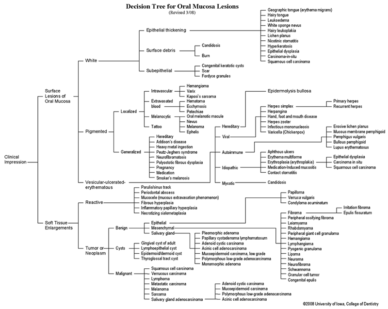 View as GIF
To view the Decision Tree for Oral Mucosal Lesions, click on one of the options shown.